|
4.
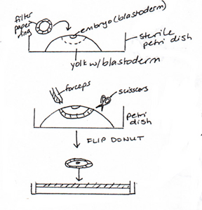
Isolate a 1- or 2-day embryo. Clean your dissecting
equipment and prepare a fresh dish of Howard's. Clean an egg
and break it into a sterile 100 mm Petri dish.
The embryo may not be
visible to the naked eye. The blastodisc is located above a
small ring of white yolk.
5.
Drop a filter paper disc around your embryo. Hold on to the
filter paper with fine forceps and cut around the ring with
your sharp scissors. Transfer the embryo to a small petri
dish with Howard's Ringer's solution.
|
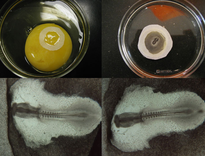
Top left - 2-day chick egg with filter paper
circle around blastodisc.
Top right - 2-day embryos isolated into Howards
Ringers
Bottom left - 2-day (HH stage 10+) embryo, dorsal
view
Bottom right - 2-day (HH stage 10+) embryo, ventral
view
|
6.
Examine both embryos. Pay particular attention to the heart
and circulation, and to the developing neural tube. Compare
the heart rate between the 2 embryos. Which side was towards
the yolk? The reddish spots on the 2-day blastodisc are the
blood islands, the sites of hematopoesis. The embryo is
covered with a clear protein layer known as the vitelline
membrane. This may start to peel away from the
embryo.
7.
For each embryo, determine the H&H
stage. How do these embryos
compare to the stained specimens?
8.
Clean your instruments well with warm water, distilled water
and 70% ethanol. Dry before returning to case. Discard the
shells and the yolk/albumin remains.
Instructor's
prep list
|

 .
.