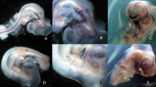
Figure 2. The expression domains
of shh in chick embryos exposed to cyclopamine.
(A-C) Three day chick embryo exposed
to 5µl of 45% HBC/PBS after 14 hours of incubation.
(D-F) Three day chick embryo exposed to 5µl of
cyclopamine/HBC complex after 14 hours of incubation. Whole
mount in situ hybridization of the embryos indicate the
presence of shh transcripts. In the control embryo (A-C),
cshh transcripts are visible along the midline, hyoid arch,
FNP, MXPs, and nasal placodes. The embryo exposed to
cyclopamine is completely lacking shh expression along its
midline (D). Although shh transcripts are present in the
hyoid arch, FNP, and nasal placodes (E-F), the level of
staining is visibly reduced in comparison to the
control.

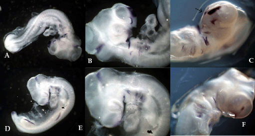
Figure 3. The expression domains
of Fgf8 in chick embryos exposed to cyclopamine.
(A-C) Three day chick embryo exposed to 5µl of 45% HBC/PBS after 14 hours of incubation. (D-F) Three day chick embryo exposed to 5µl of cyclopamine/HBC complex after 14 hours of incubation. Whole mount in situ hybridization of the embryos indicate the presence of cFgf8 transcripts. The control embryo (A-C) has clear Fgf8 expression in the limbs and the hindbrain. Within the craniofacial region, Fgf8 transcripts are located in the tip of the proboscis, the nasal placodes, and the mandibular process. High levels of Fgf8 staining appear in the hyomandibular groove. The embryo exposed to cyclopamine (D-F) also expresses Fgf8 within the limbs and hindbrain. However, there is no evidence of Fgf8 expression in the arches, mandibular process, or nasal placodes. Staining is visible along the margin of the proboscis. Fgf8 expression in chick embryos appears to be inhibited by cyclopamine only within the facial region.