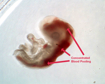
Figure 4: A chick embryo exposed to 25ug
of lead acetate. Shows severe abnormal concentrated blood
pooling throughout entire body cavity and extreme
underdevelopment.
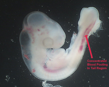
Figure 5: A chick embryo exposed to 25ug
of lead acetate. Good example of blood pooling in the tail
region which is abnormal from the control.
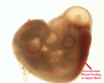
Figure 6: A chick exposed to 25ug of lead
actetate. This embryo demonstartes that lead can cause
abnormal blood pooling in the upper back of developing
embryos.
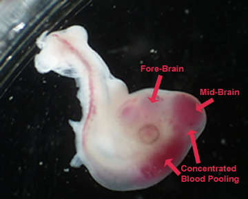
Figure 7: A chick embryo exposed to 25ug
of lead acetate. Especially good example of concentrated
pooled blood in the mid- and hind-brain regions.