|
|
 ... ...
|
Results
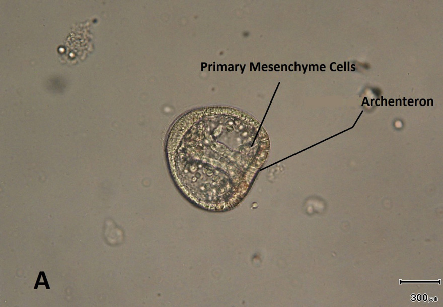
L. variegatus sea urchin gastrulation begins with the ingression of primary mesenchyme cells from the flattened epithelium at the vegetal pole of the embryos known as the vegetal plate. Following ingression of PMCs, the vegetal plate invaginates to form a cylinder called the archenteron (Fig. 1).
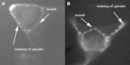
The PMCs migrate, form patterned arrays, and ultimately secrete calcium carbonate-containing skeletal rods called spicules, which can be detected using polarized light (Fig. 2) and by staining with an antibody (Ig8) to PMCs (Fig 3).
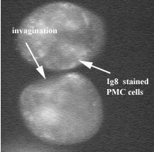
When L. variegates embryos were treated with NiCl2 from fertilization through the early gastrulation stage, the PMCs ingress and the archenteron invaginates but as gastrulation continues the PMCs do not aggregate into two ventrolateral clusters as they do in normal embryos. The Ig8 staining shows that the PMCs do not cluster or form spicules as they did in the control (Fig.4).
 |
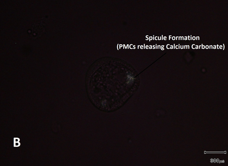 |
| Fig 1. Early Gastrulation in L. variegates embryo, the ingression of PMCs that formed the archenteron elongated across to the blastocoels to make contact with the ectoderm near the animal pole (A). Under polarized light the spicules can be seen, the PMCs start to accumulate at two sites along the ring which are called ventrolateral clusters (B). |
 |
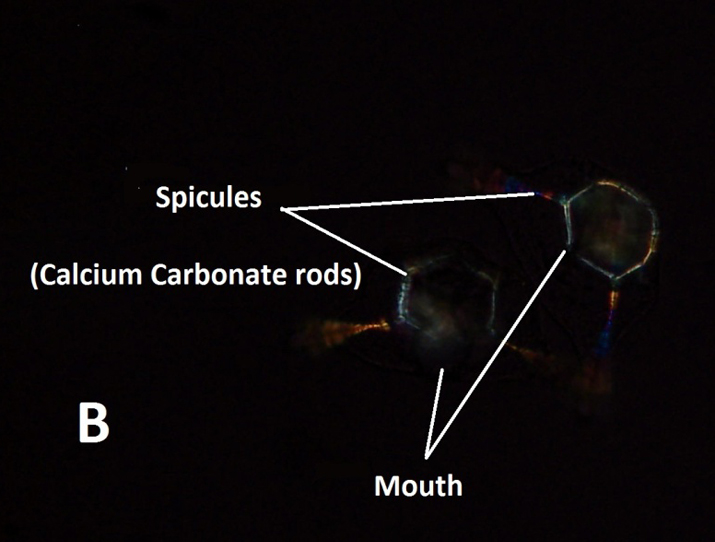 |
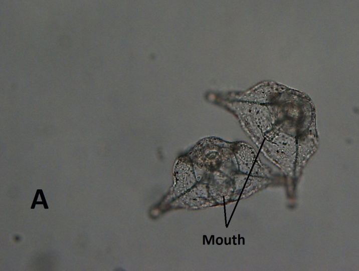
| Fig 2. Late Gastrulation in L. variegates, this can be called the prism stage (A). Calcium carbonate rods begin to form from the secretion of the PMCs in the control which gives the larval its pluteus shape (A). Under polarized light you can examine the spicule formation easily because the spicules glow (B). |
 |
 |
| Fig 3. Control pluteus larva stained with Ig8 to show the PMCs after migration and the spicules of the skeleton. (A) is a side view and (B) is a frontal view. |
Fig 4. NiCl2-treated embryos at same time after fertilization. The Ig8-stained PMCs have not clustered or formed spicules. |
|
|
