|
Results
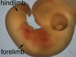
At 5 days of development, the
forelimbs and hindlimbs of the donor embryo appear as
elongated protrusions from the embryo body (fig. 4).
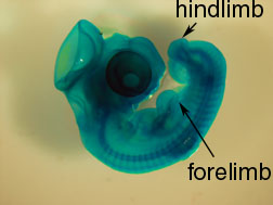
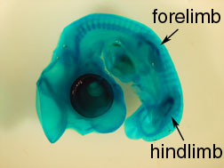
Staining with Alcian green allows visualization of cartilage
formation up to this stage in development. The 5-day old and
7-day old embryos exhibited faint cartilage at the location
of the future forelimb and hindlimbs (fig. 5a-b).
|
|
|
|
Figure 4. 5-day old donor
embryo before graft excision.
|
Figure 5a. 5-day old
embryo stained with Alcian green for cartilage
development.
|
|
|
|
|
Figure 5b. 7-day old
embryo stained with Alcian green for cartilage
development.
|
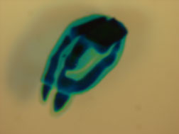
Figure 6. Hindlimb graft
derived from 7-day old donor embryo incubated on
chorioallantoic membrane for 7 days.
|
|
|
|
|
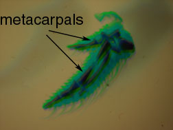
Figure 7a. Control
forelimb of 12-day old embryo. Note the upper part
of the limb is missing.
|
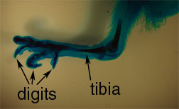
Figure 7b. Control
hindlimb of 12-day old embryo.
|
|
|
|
|
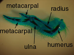
Figure 7c. Control
forelimb of 14-day old embryo.
|
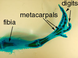
Figure 7d. Control
hindlimb of 14-day old embryo.
|
Of the 12 grafts attempted from
5-day old donor embryos in one experiment, none were
successful. Of the 3 grafts derived from the 7-day old donor
embryos, one was successful. Many of the embryos died from
infection during the 7-day incubation period, as evidenced
by the yellow-green appearance and odor of the egg contents.
Of the embryos that survived, the grafts were difficult or
impossible to locate, possibly because the grafts were
concealed by membrane or lost in the liquid environment
below the membrane.
The only successful graft was a
hindlimb derived from a 7-day old embryo. After the 7-day
incubation period on the chorioallantoic membrane, the graft
showed considerable cartilage development (compare fig. 5b,
6). However, only the most distal structures developed,
compaared with the intact 7-day control embryos incubated
for 7 days (fig. 7d).
Though no grafts derived from
5-day old embryos or forelimbs from 7-day old embryos were
successful, it is worthwhile to compare limb development in
the controls that were prepared. After 7 days of incubation,
the embryos were 12 and 14 days old, respectively. There is
extensive cartilage and bone formation in both groups.
Distinct joints can be discerned, as well as clearly
distinguished digits in the forelimbs and hindlimbs (fig.
7a-d).
|