|
|

|
Results
There
were three different trials run in order to see if we could
get successful neural crest cell migration. All of our
control wells showed a large amount of neuron
differentiation, with the neurite formation quite visible
after staining with anti-neurofilament antibody. Most of the
neurons that formed in the control wells were normal in
appearance with only one main neurite branch. The two wells
containing 1 mM Pb-Acetate showed a few neurons that had
differentiated. These neurons seemed to have one main
neurite with multiple branches. The two wells with 0.1 mM
Pb- Acetate had a considerable number of differentiated
neurons. In one well, there were two large webs of neurons,
most of which had multiple branches coming off of each
neuron axon body (see figures below).
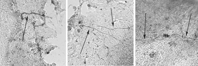
Control neural crest
cultures
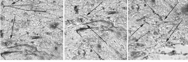
Neural crest cultures
treated with lead acetate
|
|