|
|
|
Background
In the early development of
every member of the chordate phylum, somites arise from the
dorsal mesoderm. These segments increase in number as the
organism grows and are a useful determination of the
embryo's age. Somite segmentation has been shown to be
dependent on specific cell-cell and cell-matrix interactions
during embryogenesis, and these interactions are thought to
be regulated by the expression of pattern-forming genes
(Barnes et. al., 1996). The formation and patterning of the
axial vertebral skeleton is the target of a number of
teratogenic agents during embryonic development.
The Pax-1 gene has been
shown to be an important regulator of axial skeletal
patterning and is specifically implicated in the formation
and maturation of the distinct somite sclerotome that gives
rise to the axial skeleton as vertebrae, intervertebral
discs, ribs, and neural arches (Smith & Tuan, 1995).
Researchers determined that Pax-1 gene expression is
critical to the normal development of somites in chicks, and
has been found to play a similar role in the somites of rats
and mice (Vorhees, 1987). The Pax-1 gene is expressed in
newly formed epithelial somites as well as sclerotomal cells
and remains a target of teratogens that generate anomalies
of the axial skeleton.
Valproic acid is an anticonvulsant drug used to treat
epilepsy. Since its release for human use in 1967, it has
been found to cause a number of congenital birth defects in
the children of women who use the drug. Such defects include
facial malformations and skeletal abnormalities (Lammer et.
al., 1987). Since nearly 1 out of 200 pregnant women is
epileptic and a candidate for valproic acid treatment,
researchers have aimed to determine which gene was affected
by the presence of valproic acid. Recent studies have
concluded that the mechanism by which valproic acid causes
these birth defects is by down-regulating the expression of
the Pax-1 gene (Lammer et. al., 1987). The molecular pathway
of valproic acid, as in a large number of pharmaceutical
teratogens, currently remains unknown. However one theory
suggests that valproic acid acts via a deficiency in the
vitamin folic acid, as folic acid deficiency is linked to an
increased incidence of neural tube defects in humans
(Barnes, et. al., 1996). Specifically, when tested on
chicken embryos, valproic acid affected Pax-1 expression,
causing the disorganization or fusion of somites (Barnes et.
al., 1996). Other studies showed that rats treated with
valproic acid demonstrated similar results of fused ribs,
missing vertebrae, and kinky tails (Vorhees, 1987). This
research demonstrates that the Pax-1 gene is crucial to the
normal development of somites in chickens, mice, and rats,
and the down-regulation of Pax-1 due to valproic acid will
result in somite malformations in all of these organisms
(Vorhees, 1987).

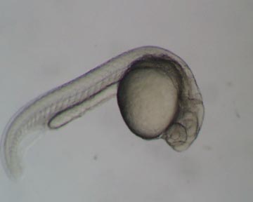
Figure 1. Danio rerio embryo at 28 hours with normal
development of somites.
© Cebra-Thomas,
2001
Last Modified: 31 May
2001
[Lab
Protocols
| Students
| Cebra-Thomas
| Course
| Links
]
|
|
