|
|
|
Upon
fixing and staining of our cells, we observed definite
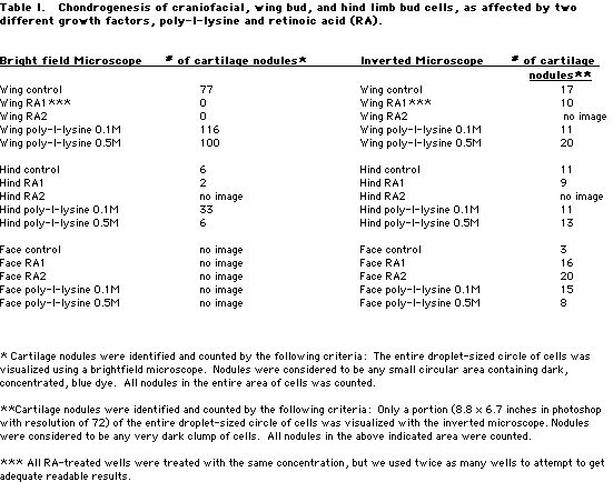
cartilage formation in both the wing and leg controls
(Figure 1 B1 & Figure 2 C1, table 1). The face cells
never adhered properly to the base of the wells. Therefore,
when the growth media was pulled out in order to add the
paraformaldehyde, the cells unavoidably came out with the
media. The hind and wing cells did not come out with the
media. They remained in a circular shape on the bottom of
the wells in their adhered positions. We took two kinds of
pictures. The first was a set of images taken with the
inverted scope prior to pulling out the growth media. By
doing this, we were hoping to get pictures of chondrogenesis
in the face cells without disturbing them. However, we still
were not able to visualize the face controls fully because
the cells had been scattered before we could take the
pictures. We did see three cartilage nodules in the face
control, so we know that chondrogenesis did occur (Figure 1
A1). We just do not know how much.
After treatment of each type
of cells with retinoic acid (RA), we observed some
interesting, but unfortunate occurrences. The RA seemed to
settle out of solution and remain on the bottom of the wells
in 8 out of the 12 RA-treated wells. This occurrence made it
extremely difficult to view the chondrogenesis that may have
taken place. We used RA that had been made up for a previous
experiment. For future experiments, the RA should be made up
fresh, so as to potentially avoid this problem. We took
pictures of the wells that were free enough of RA so that
they could be visualized (Figure 1 A2, B2, & C2).
Generally, we were not able to conclude that RA inhibited
chondrogenesis in hind limb buds and stimulated it in wing
buds as literature suggests (Figure 2 B2, C2) (Smith 1998).
The face results were again indiscernible in the bright
field microscope after staining. Prior to fixing and
staining, however, the pictures from the inverted scope
showed slightly more chondrogenesis than was seen for the
hind limb cells

|
|