|
Prep
list
|
Drosophila 3rd
instar larvae
sterile petri
dishes
sterile
forceps
tungsten
microneedles
pulled Pasteur
pipettes
sterile glass
pipettes
paintbrushes
|
pipettors
depression
slides
sterile insect
saline
70% and 95%
ethanol
Schneider's insect medium
with 10% fetal calf serum
ecdysone solution (1mg/mL
in ethanol)
Gentamicin reagent
solution (50 mg/mL)
|
Procedure
1. Use sterile technique
throughout this experiment.
2. Prepare four 35-mm culture
dishes by adding 2 mL of serum-supplemented Schneider's
insect medium to each dish and 2 uL Gentamicin.
a. To one plate, add 8 uL of
ecdysone solution (1 mg powder per mL 95% ethanol). Final
concentration of ecdysone 4 ug/ml.
b. To the second plate, add 5 uL
of ecdysone solution.
c. Add 2 uL ecdysone solution to
the third dish.
d. The fourth control plate should
receive 5 uL of 95% ethanol.
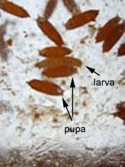
3. Obtain a large third-instar
maggot from container (fig. 1, 2) and rinse it clean of
debris by soaking it in several drops of saline on
depression slide or other small container. Place container
over ice to anesthetize and slow larvae
responses.
4. Transfer larvae to several
drops of cold saline solution on a clean glass plate (fig.
3).
5. Under a dissecting microscope
remove as many imaginal discs as possible.
a. Grasp the head of larva just
behind the mouth parts with one pair of forceps.
b. Grasp the middle of the larva
with the other pair and gently pull the body away from the
head.
c. The imaginal discs will be
concentrated in the anterior third of the body, attached to
the tracheoles or gut (fig. 4).
6. Dissect the discs free of
adhering tissue (fig. 5); the wings and legs discs are the
largest and generally evert most drastically.
7. Dissect enough larvae to obtain
20 discs.
8. Transfer 4 discs into each dish
using pulled Pasteur pipettes or tungsten
microneedles.
9. Examine and photograph each
disc.
10. Photograph and examine discs
at two hours and 24 hours.
11. Allow discs to incubate at
room temperature during development.

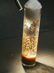
Figure 1. Drosophila 3rd instar larvae in
tube of fly medium.
|

Figure 2. Drosophila 3rd
instar larva (close-up).
|

|

|
|
Figure 3. Magnified photograph of 3rd instar
Drosophila larva as viewed through
dissecting microscope.
|
Figure 4. Drosophila larva prior to
removal of imaginal discs from larval body.
|
|

Figure 5. Imaginal discs (im) free of debris and
ready for transfer to culture dishes.
|
|
|
|
|
|


![]()
![]()
![]()