|
|
Objective Intro Materials Procedure Results Discussion Lit. Cited Prep Sheet |
||
|
|
|
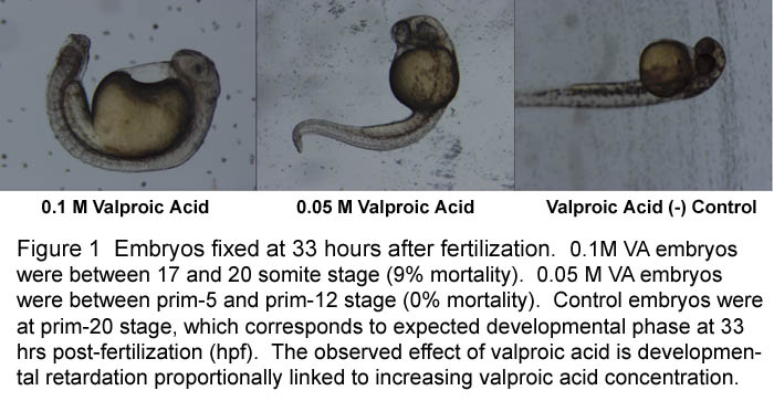
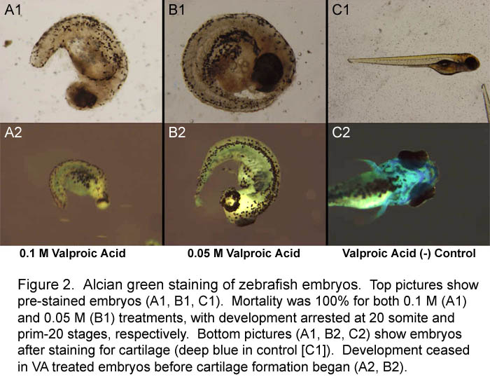
Results The embryos subjected to valproic acid (VA) exhibited observable developmental retardation as a function of increasing VA concentration, as well as noticeable tail malformation when compared to control embryos (Figure 1). Mortality rates were low for both concentrations (0.05 M- 0% / 0.1 M- 9% / control- 0%). All embryos for F6 staining were fixed at approximately 33 hours after fertilization. When fixed, control embryos were at the corresponding prim-20 stage of development (Kimmel, 1995). Embryos in 0.05 M VA were between the prim-5 and prim-12 stages (equivalent to 24-28 hrs after fertilization). Embryos in 0.1 M VA were between the 17-somite and 20-somite stages (equivalent to 17.5-19 hrs after fertilization). Staining for somite boundaries was unsuccessful, presumably due a decrease in the binding affinity of the primary F6 antibody or the secondary HRP antibody, both of which came from old stock solutions. Embryos that were to be stained with alcian green did not
survive long enough to develop cartilage. Precartilage
condensation begins at approximately 60 hrs after
fertilization (Kimmel, 1995). Mortality rates for both 0.05
M VA and 0.1 M VA embryos were 100%. Development was
arrested at approximately the prim-20 stage (33 hr
equivalent) for 0.05 M VA embryos and 20-somite stage (19 hr
equivalent) 0.1 M VA embryos (Figure 2).
Control embryos were at the 4 day corresponding
developmental stage and exhibited significant pre-bone
formation.
Last Modified: 14 July 2014 [Lab Protocols | Students | Cebra-Thomas | Course | Links ] |