![]()
![]()
![]() Objectives Introduction
Procedure
Results Figure Discussion
Acknowledgements
and References
Objectives Introduction
Procedure
Results Figure Discussion
Acknowledgements
and References ![]()
|
|
|
|
|
|
|
|
|
|
|
|
|
|
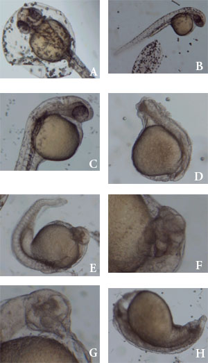
Figure 1. The embryos were all photographed approximately 30 hours after fertilization. A and B are normally developed embryos in the ZF control medium, staged at 42 hpf. C has retarded development (~31 hpf) but shows no deformities in the 1% ethanol control. D, in the 3% ethanol control, lacks somites and has a poorly developed tail that has not extended from the yolk; by contrast the head extends off the yolk but is also underdeveloped. E, (20 mg/ml cyclopamine solution,) has organized somites but a bent posterior section, underdeveloped eyes (compare with C), and a deformed head, and exhibits retarded growth (~31 hpf). F and G show eye deformities in the same solution. F has two eyes that are anteriorly displaced while G lacks eyes but has a single lens (in the head just above the highest part of the yolk). H, in the 60 mg/ml cyclopamine solution, has retarded growth (~17 hpf) has a distended yolk, lacks a posterior and distinguishable somites and has a malformed head. |