|
|
Culturing skin from eight day chicken embryos: the development of feather buds |
Erin Betters, Katy Lewis, Katie Crawford Swarthmore College
|


Introduction:
Within chick embryos, interactions between the epidermis and dermis of the skin result in the formation of preliminary feather buds, and later, specific feather types or cutaneous structures (Gilbert, 2003). The outer layer of skin, the epidermis, is formed from the ectoderm of the developing embryo; it covers the embryo following the development of the neural tube (Gilbert, 2003). After neurulation, the preliminary epidermis undergoes a series of cell divisions and dif-ferentiations to form the skin. The final product consists of the basal layer, or germinal epithelium, the spinous layer, the granular layer, and cells known as keratinocytes (Gilbert, 2003).
Altogether, the cells of the epidermis are connected and
form an epithelium, or sheet of cells (Gilbert, 2003). Under
the epidermis lies the dermis, which has its origins from
the meso-derm and is a mesenchymal layer of “loosely”
associated cells (Gilbert, 2003). The epidermis signals to
the dermis through the production of sonic hedgehog and
transforming growth factor-beta (SHH, TGF-beta) proteins.
Once the dermis receives these factors its cells aggregate,
and in turn signal back to the epidermis, causing certain
genes to be activated (Gilbert, 2003). As a re-sult,
different cutaneous structures are formed, based on the
location of the dermis (Gilbert, 2003). Of particular note,
such interactions between the epidermis and dermis layers
lead to the initial development of feather buds. In the
early stages, parts of the epidermis thicken and form
epidermal palcodes, under which the dermal cells aggregate
to form dermal papilla – a small projection of tissue
at the base of the feather (Bellairs et al., 1998). In time,
feather buds appear as these structures elongate, forming
the basis from which feathers will be formed (Bellairs et
al., 1998)(figure 2C).
Understanding that the development of the skin can occur
autonomously from the rest of the organism, specifically in
culture, has proven vital to the development of techniques
to treat burn injuries, as well as factored into the
creation of artificial skin such as Apligraf (OI, 2003;
Scott, 2000). Apligraf, composed of a collagen matrix,
fibroblast cells (which contribute to the dermis), and
keratinocytes, has been used to treat both diabetic and
venous ulcers. (OI, 2003; Cebra-Thomas, 2004). If the
complexities of skin formation can be understood, strides
could be made in the development of skin grafts, allowing
for greater availability to patients. In addition, greater
understanding of the chemical signals between the dermal and
epidermal layers could be used to design treatments for burn
patients whose skin layers have been damaged, helping the
patient’s own skin regenerate rather than depending on
whole skin grafts.
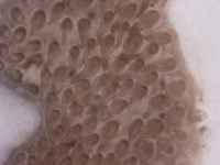
This experiment seeks to demonstrate that the skin of
eight-day-old chick embryos, if re-moved, can continue to
develop to the feather bud stage in culture(figure
2C). We expect that, as in past experiments, this
development will be successful to the point of late feather
bud development, but will arrest before any actual feather
development takes place (as it would in-ovo) (Cebra-Thomas,
2004).