|
|
AC
|
LF
|
LT
|
3/2/04 (Day 1)
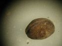
Figure 4. Stage 15 donor before operation -
donors were younger than hosts
|
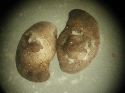
Figure 5. AC 1 (left) and 2 (right). Grafts seem
to be adhering well.
|
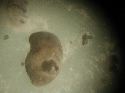
Figure 6. LF - example of one of three
grafts
|
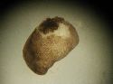
Figure 7. LT - example of one of five grafts
|
|
3/3/04 (Day 2)
|
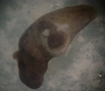
Figure 8. AC 1 - ragged edge around graft
visible
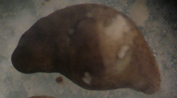
Figure 9. AC 2 - graft nearly healed
|
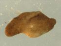
Figure 10. LF - only graft of three; has healed
flawlessly
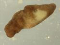
Figure 11. LF - has rejected its graft (white
area is the wound)
|
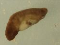
Figure 12. Only remaining graft; graft is dark
brown patch in center of white wound
|
|
3/4/04 (Day 3)
|
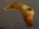
Figure 13. Nearly healed (AC 1; AC 2 dead)
|
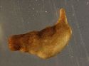
Figure 14. Perfectly healed
|
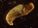
Figure 15. Graft not securely attached
|
|
3/5/04 (Day 4)
|
|
|
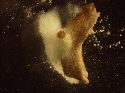
Figure 16. Graft rejected, embryo dead
|
|
3/6/04 (Day 5)
|
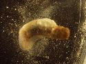
Figure 17. Wound on embryo has enlarged,
spilling inner contents
|
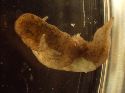
Figure 18. Successful graft has differentiated
somewhat, host is alive and moving
|

Figure 19. Control (very healthy; normal
development)
|